ANALYSIS OF OCT-A PERFORMED AMONG NAION PATIENTS VERSUS OTHER CAUSES

Article Sidebar
- Non-arteritic anterior ischemic optic neuropathy, papil atrophy, optical coherence tomography angiography
Abstract
Introduction:Non-Arteritic Anterior Ischemic Optic Neuropathy (NAION) is the type of optic neuropathy with crowding structural of microcirculation on optic nerve head. The vascular factor has the role on its pathophysiology. The aim of this study to analyse OCT-A among NAION patients compared to patients of papil atrophy with other causes.
Methods:A retrospective case control study held from medical record of patients. Two group diagnosis of NAION and papil atrophy due to other causes who performed OCT-A in Dr. Kariadi Hospital Semarang. The analysis of the study including vessel perfusion density (VPD) and flux index (FI) among two groups. The bivariate analysis is using Independent-T test.
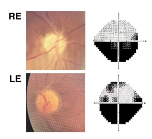
Results:Totally 40 patients consisted of 20 NAION (12 bilateral eyes) and 20 papil atrophy with other causes (15 bilateral eyes). Age over 50 years old were 19 NAION (95%) and 17 other causes (85%). The results found there is statistically significant difference between VPD among NAION and other causes in average, superior, and nasal (p value <0.05, respectively 0.008, 0.008, 0.030), but there is no significant difference between FI among NAION and other causes both average, inferior, superior, nasal, and temporal (p value > 0.05). Mean VPD both average, inferior, superior, nasal and temporal in NAION were lower than other causes (respectively 39,98±3,69, 38,57±6,01, 36,77±5,25, 40,78±4,06, 43,91±3,42).
Conclusion:This study found there is statistically significant difference between VPD among NAION and other causes in average, superior, and nasal. VPD in NAION were lower compared to other causes.
Full text article
References
American Academy of Ophthalmology. Neuro-Ophthalmology. In: Bhatti MT, et al., editors. Basic and Clinical Science Course. Section 5. San Fransisco: American Academy of Ophthalmology. 2022. p. 153-155.
Ling L, et al. Optical Coherence Tomography Angiography Assesment of the Peripapillary Vessel Density and Structure in Patients with Nonarteritic Anterior Ischemic Optic Neuropathy: A Meta-Analysis. Biomed Research Journal. 2020. 1-11. Available from: https://doi.org/10.1155/2020/1359120.
Hayreh SS, Zimmerman MB. Nonarteritic Anterior Ischemic Optic Neuropathy. Natural History of Visual Outcome. Ophthalmology. 2008 Feb;115(2):298-305.
Sharma S, Kwan S, Fallano KA, Wang J, Miller NR, Subramanian PS. Comparison of Visual Outcomes of Nonarteritic Anterior Ischemic Optic Neuropathy in Patients with and without Diabetes Mellitus. Ophthalmology. 2017 Apr 1;124(4):450–5.
Pinna A, Solinas G, Masia C, Zinellu A, Carru C, Carta A. Glucose-6-phosphate dehydrogenase (G6PD) deficiency in nonarteritic anterior ischemic optic Neuropathy in a sardinian population, Italy. Investigative Ophthalmology and Visual Science. 2008 Apr;49(4):1328–32.
Yao F, Wan P, Su Y, Liao R, Zhu W. Impaired systemic vascular endothelial function in patients with non-arteritic anterior ischaemic optic neuropathy. Graefe’s Archive for Clinical and Experimental Ophthalmology. 2016 May 1;254(5):977–81.
Giambene B, et al. Evaluation of traditional and emerging cardiovascular risk factors in patients with non-arteritic anterior ischemic optic neuropathy: A case-control study. Graefe’s Archive for Clinical and Experimental Ophthalmology. 2009 May;247(5):693–7.
Sharma S, et al. Optical coherence tomography angiography in acute non-arteritic anterior ischaemic optic neuropathy. British Journal of Ophthalmology. 2017 Aug 1;101(8):1045–51.
Gandhi U, et al. Optical coherence tomography angiography in acute unilateral nonarteritic anterior ischemic optic neuropathy: A comparison with the fellow eye and with eyes with papilledema. Indian Journal of Ophthalmology. 2018 Aug;66(8):1144-48.
Jia Y, et al. Optical coherence tomography angiography of optic disc perfusion in glaucoma. Ophthalmology. 2014 Jul. 121(7):1322-32.
Hata M, et al. Structural and Functional Analyses in Nonaretritic Anterior Ischemic Optic Neuropathy: Optical Coherence Tomography Angiopgraphy Study. J Neuroophthalmol. 2017 Jun;37(2):140-8.
Song Y, Min JY, Mao L, Gong YY. Microvasculature dropout detected by the optical coherence tomography angiography in nonarteritic anterior ischemic optic neuropathy. Lasers Surg Med. 2018 Maret;50(3):194-201.
Liu CH, Kao LY, Sun MH, Wu WC, Chen HSL. Retinal vessel density in optical coherence tomography angiography in optic atrophy after nonarteritic anterior ischemic optic neuropathy. J Ophthalmol. 2017. 2017:9632647. Avalaible from: https://doi.org/10.1155/2017/9632647.
Olver JM, Spalton DJ, McCartney AC. Microvasular study of the retolaminar optic nerve in man: the possible significance in anterior ischemic optic neuropathy. Eye (Lond). 1990;4(Pt 1):7-24.
Balducci N, et al. Optical coherence tomography angiography in acute arteritic and non-arteritic anterior ischemic optic neuropathy. Graefe’s Archive for Clinical and Experimental Ophthalmology. 2017 Nov; 255(11):2255-2261.
Rebolleda G, Diez-Alvarez L, Garcia Y, et al. Reduction of peripapillary vessel density by optical coherence tomography angiography from the acute to the athropic stage in non-arteritic ischaemic optic neuropathy. Ophthalmologica. 2018;240(4):191-199.
Authors
Copyright (c) 2025 Usamah Haidar, Riski Prihatningtias

This work is licensed under a Creative Commons Attribution-NonCommercial-ShareAlike 4.0 International License.