The Role of Optical Coherence Tomography Angiography in Non-Arteritic Ischemic Optic Neuropathy: A Case Report

Article Sidebar
- non-arteritic ischemic optic neuropathy, visual field defect, optical coherence tomography angiography
Abstract
Introduction: Non-arteritic ischemic optic neuropathy (NAION) is the second most common optic neuropathy in adults. Since circulatory insufficiency is presumed to have a role in pathogenesis of NAION, optical coherence tomography angiography (OCTA) is suggested as a tool in assessing NAION patients, while optical coherence tomography (OCT) can detect structural changes. This case report aims to describe the congruity of visual field defect with OCT and OCTA result in a case of bilateral NAION, highlighting the role of both OCT and OCTA in NAION.
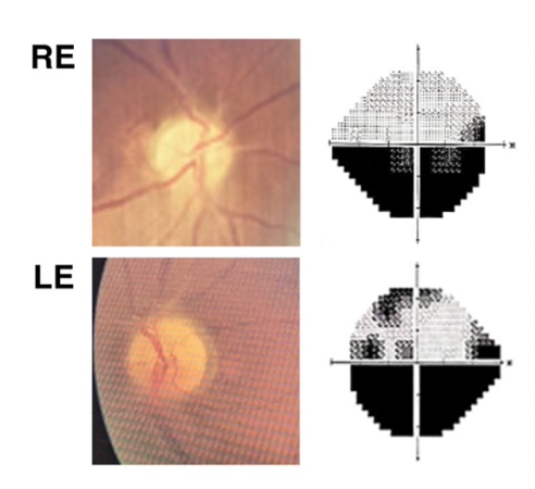
Case Report: A 52-years-old male came due to painless, progressive blurry vision in both eyes, especially in lower visual field, for the past 7 months. Humphrey visual field examination revealed inferior visual field defects in both eyes. OCTA showed reduced retinal perfusion in the superior part bilaterally. OCT revealed ganglion cell loss in the superior part of his right eye and almost all parts of his left eye. Retinal nerve fiber layer (RNFL) thinning was found in the superior part bilaterally.
Discussion: In this case, the congruity of visual field defect, reduced perfusion, ganglion cell-inner plexiform layer (GCIPL), and RNFL thinning portrayed the connection of hypoperfusion as the presumed underlying mechanism of NAION, neuron loss as the result of the hypoperfusion, and the visual field loss as the presenting symptom in NAION.
Conclusion: This finding demonstrates the role of OCT-A and OCT in diagnosing and monitoring progression in patients with NAION.OCT-A is a useful tool to evaluate microvascular changes, while OCT can be used to evaluate RNFL and GCIPL thinning.
Full text article
References
Moghimi S, Afzali M, Akbari M, Ebrahimi KB, Khodabande A, Yazdani-Abyaneh AR, et al. Crowded optic nerve head evaluation with optical coherence tomography in anterior ischemic optic neuropathy. Eye (Basingstoke). 2017 Aug 1;31(8):1191–8.
Berry S, Lin W v., Sadaka A, Lee AG. Nonarteritic anterior ischemic optic neuropathy: Cause, effect, and management. Vol. 9, Eye and Brain. Dove Medical Press Ltd.; 2017. p. 23–8.
Lee JY, Park KA, Oh SY. Prevalence and incidence of non-arteritic anterior ischaemic optic neuropathy in South Korea: A nationwide population-based study. British Journal of Ophthalmology. 2018 Jul 1;102(7):936–41.
Zulkarnaen M, Nusanti S, Sidik M, Rachman A. A Five-Year Data in Descriptive Study of Systemic and Ocular Risk Factors of Non-arteritic Anterior Ischemic Optic Neuropathy (NAION) and The Correlation to The Clinical Condition. Vol. 47, Ophthalmol Ina. 2021.
Rougier MB, Gattoussi S, Le-Goff M, Korobelnik JF. OCT angiography analysis in acute non-arteritic anterior ischemic optic neuropathy: The importance of segmentation. European Journal of Ophthalmology. 2021 Nov 1;31(6):3471–5.
Sharma S, Ang M, Najjar RP, Sng C, Cheung CY, Rukmini A v., et al. Optical coherence tomography angiography in acute non-arteritic anterior ischaemic optic neuropathy. British Journal of Ophthalmology. 2017 Aug 1;101(8):1045–51.
Liu B, Yu Y, Liu W, Deng T, Xiang D. Risk Factors for Non-arteritic Anterior Ischemic Optic Neuropathy: A Large Scale Meta-Analysis. Vol. 8, Frontiers in Medicine. Frontiers Media S.A.; 2021.
Miller NR, Arnold AC. Current concepts in the diagnosis, pathogenesis and management of nonarteritic anterior ischaemic optic neuropathy. Vol. 29, Eye (Basingstoke). Nature Publishing Group; 2015. p. 65–79.
Pujari A, Bhaskaran K, Sharma P, Singh P, Phuljhele S, Saxena R, et al. Optical coherence tomography angiography in neuro-ophthalmology: Current clinical role and future perspectives. Vol. 66, Survey of Ophthalmology. Elsevier Inc.; 2021. p. 471–81.
Mayes EW, Cole ED, Dang S, Novais EA, Vuong L, Mendoza-Santiesteban C, et al. Optical coherence tomography angiography in nonarteritic anterior ischemic optic neuropathy. Journal of Neuro-Ophthalmology. 2017 Mar 15;37(4):358–64.
Contreras I, Noval S, Rebolleda G, Muñoz-Negrete FJ. Follow-up of Nonarteritic Anterior Ischemic Optic Neuropathy with Optical Coherence Tomography. Ophthalmology. 2007;114(12).
Rebolleda G, Diez-Alvarez L, Casado A, Sánchez-Sánchez C, de Dompablo E, González-López JJ, et al. OCT: New perspectives in neuro-ophthalmology. Vol. 29, Saudi Journal of Ophthalmology. Elsevier; 2015. p. 9–25.
Akbari M, Abdi P, Fard MA, Afzali M, Ameri A, Yazdani-Abyaneh A, et al. Retinal ganglion cell loss precedes retinal nerve fiber thinning in nonar teritic anterior ischemic optic neuropathy. Journal of Neuro-Ophthalmology. 2016;36(2):141–6.
de Dompablo E, García-Montesinos J, Muñoz-Negrete FJ, Rebolleda G. Ganglion cell analysis at acute episode of nonarteritic anterior ischemic optic neuropathy to predict irreversible damage. A prospective study. Graefe’s Archive for Clinical and Experimental Ophthalmology. 2016 Sep 1;254(9):1793–800.
Moghimi S, Afzali M, Akbari M, Ebrahimi KB, Khodabande A, Yazdani-Abyaneh AR, et al. Crowded optic nerve head evaluation with optical coherence tomography in anterior ischemic optic neuropathy. Eye (Basingstoke). 2017 Aug 1;31(8):1191–8.
Giordano M, Montorio D, Concilio M, Morra VB, Cennamo G. Peripapillary vascular density in resolved non-arteritic anterior ischemic optic neuropathy: colocalization between structural and vascular parameters. Neurological Sciences. 2021 Nov 1;42(11):4723–5.
Higashiyama T, Ichiyama Y, Muraki S, Nishida Y, Ohji M. Optical Coherence Tomography Angiography in a Patient with Optic Atrophy After Non-arteritic Anterior Ischaemic Optic Neuropathy. Neuro-Ophthalmology. 2016 May 3;40(3):146–9.
Sharma S, Ang M, Najjar RP, Sng C, Cheung CY, Rukmini A v., et al. Optical coherence tomography angiography in acute non-arteritic anterior ischaemic optic neuropathy. British Journal of Ophthalmology. 2017 Aug 1;101(8):1045–51.
Rebolleda G, Diéz-Álvarez L, Garciá Marín Y, de Juan V, Munõz-Negrete FJ. Reduction of peripapillary vessel density by optical coherence tomography angiography from the acute to the atrophic stage in non-arteritic anterior ischaemic optic neuropathy. Ophthalmologica. 2018 Nov 1;240(4):191–9.
Huang HM, Wu PC, Kuo HK, Chen YJ, Poon LYC. Natural history and visual outcome of nonarteritic anterior ischemic optic neuropathy in Southern Taiwan: a pilot study. International Ophthalmology. 2020 Oct 1;40(10):2667–76.
Authors
Copyright (c) 2022 Syntia Nusanti, Brigitta Marcia Budihardja, Lazuardiah Anandi, Rona Ali Badjrai

This work is licensed under a Creative Commons Attribution-NonCommercial-ShareAlike 4.0 International License.