Can Optical Coherence Tomography Angiography be a Useful Adjunctive Diagnostic Tool in Patient with Non Arteritic Ischaemic Optic Neuropathy? Poster Presentation - Case Series - Resident
Abstract
Abstract
Introduction : Perfusion deficiency of the optic nerve head is presumed to have an immense role in the pathogenesis of non-arteritic ischaemic optic neuropathy (NAION). Optical coherence tomography angiography (OCTA) is a non-invasive method that can assess posterior pole vasculature at multiple levels. Thus, these case series want to compare the OCTA in the acute (before four weeks) and chronic phases (after six weeks) of NAION with their fellow unaffected eyes to give an overview of vasculature changes in NAION.
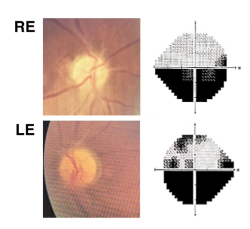
Case Illustration : This case series consists of four patients with NAION. Half of them came with a sudden painless blurry vision, and the remaining presented with an abrupt visual field defect. All of them have diabetes mellitus and hypertension. None of them have superimposed vascular pathology. The first OCTA was taken within seven days of the first symptoms, and the second OCTA was taken more than six weeks after.
Discussion : The high percentage of systemic vascular risk factors and the acute onset supported the vascular occlusive hypothesis in the pathogenesis of NAION. Meanwhile, the OCTA provides an actual overview of the vasculature changes. Microvasculature is more likely to expand in the acute phase, and a more significant expansion was found in greater optic disc edema. On the other hand, microvascular tend to attenuate in the chronic phase, representing the pruning of capillaries. A swept-source OCTA with deeper penetration might give a better peripapillary choriocapillaris image in the acute phase.
Conclusion : OCTA is a promising adjunctive tool for diagnosing and monitoring progression in patients with NAION.
Full text article
References
(-)
Authors

This work is licensed under a Creative Commons Attribution-NonCommercial-ShareAlike 4.0 International License.


