
Changes in Pattern and Multifocal Electroretinogram in Tuberculosis Patient with Ethambutol Therapy

Article Sidebar
- electroretinography, ethambutol, neuropathy, pattern ERG, multifocal ERG
Abstract
Introduction: Tuberculosis (TB) is a world health problem, especially in Indonesia as the third biggest country for new emerging TB patients. Ethambutol is one of the standard therapies to treat TB patients in Indonesia. Ethambutol has a side effect called ethambutol optic neuropathy which is hard to diagnose due to normal fundus appearance in most cases and therefore often detected late. Early detection is necessary so that permanent damage can be prevented. Examination pattern electroretinography (pERG) and multifocal electroretinography (mfERG) have the advantage to detect and confirm ocular toxicity by ethambutol after the clinical problem had emerged. It is not yet known neither pERG nor mfERG could detect any changes to detect ethambutol ocular toxicity before the clinical problem emerged.
Methods: This study was a prospective clinical trial with 40 eyes samples and analyzed with paired t and Wilcoxon tests. The ocular examination was conducted using the Snellen chart, HRR Richmond Plates, Pelli Robson, pERG, and mfERG in tuberculosis category 1 patient with 2 months follow-up.
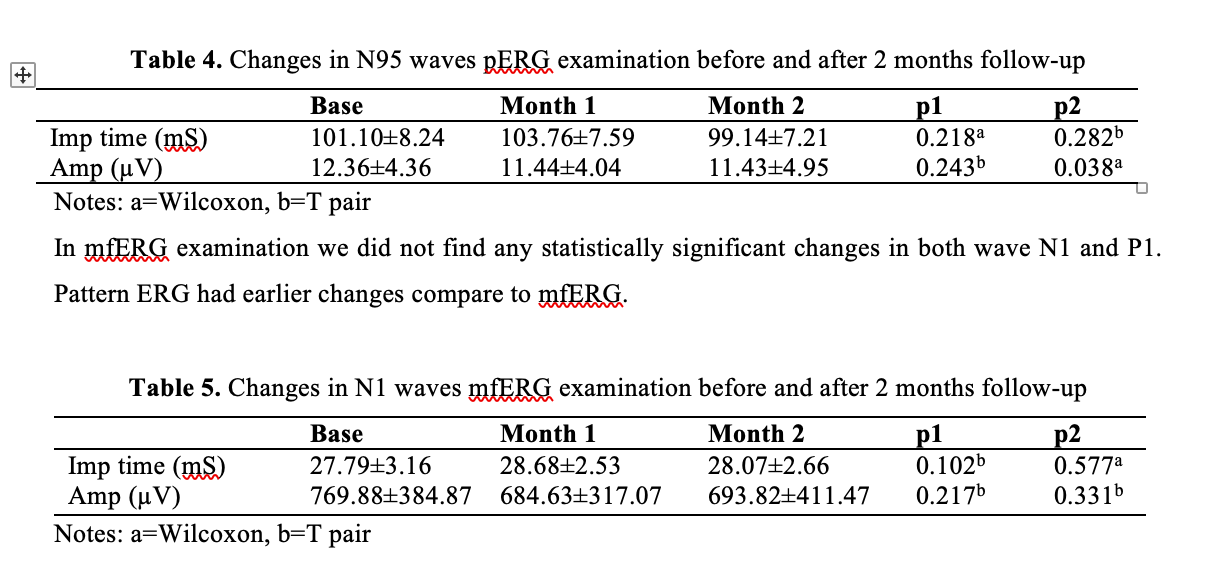
Result: Visual acuity, color, and contrast sensitivity were normal in all patients for 2 months follow-up period. In pERG examination, the mean implicit time wave P50 was shortened by -1.27± 4.71 mS (p=0.049), and the mean amplitude wave N95 was reduced by -0.93± 4.49 ?V (p=0.038). Both were statistically significant. In the mfERG examination, we did not find any statistically significant changes in both wave N1 and P1.
Conclusion: Changes in pattern ERG presented earlier compared to mfERG after ethambutol therapy for 2 months.
Full text article
References
WHO. Global tuberculosis report 2016. A new era of global TB monitoring. 2016;2(3):5-15.
Kementrian Kesehatan Republik Indonesia. Pedoman Nasional Pengendalian Tuberkulosis. Indonesia; 2014.
Junita P, Sidik M. Karakteristik Persepsi Warna dan Sensitivitas Kontras pada Pengguna Etambutol. Universitas Indonesia; 2009.
Chan H, Ng Y, Chu P. Applications of the Multifocal Electroretinogram in the Detection of Glaucoma. Clinical & Experimental Optometry. 2011;94(3):247-.
Barron G, Tepper L, Lovine G. Ocular Toxicity from Ethambutol. American Journal of Ophthalmology. 1974;77(2):256-.
Bach M, Hoffmann M. Update on the Pattern Electroretinogram in Glaucoma. Optometry and Vision Science?: Official Publication of the American Academy of Optometry. 2008;85(6):386-.
Hood D, Odel J, Chen C, Winn B. The Multifocal Electroretinogram. Journal of Neuro ophthalmology: The Official Journal of the North American Neuro-Ophthalmology Society. 2003;23(3):225-.
Jalali S, Holder G, Ram L, Vedantam V. Visual Electrophysiology inThe Clinic: A Basic Guide to Recording and Interpretation. All Indian Ophthalmological Society CME Series 17. 2009;5-6:5-15.
Balasopoulou A, ?okkinos P, Pagoulatos D, Plotas P, Makri OE, Georgakopoulos CD, et al. Symposium Recent advances and challenges in the management of retinoblastoma Globe saving Treatments. BMC Ophthalmology. 2017;17(1):1.
Geyer HL, Herskovitz S, Slamovits TL, Schaumburg HH. Optochiasmatic and peripheral neuropathy due to ethambutol overtreatment. Journal of Neuro-Ophthalmology. 2014;34(3):257–8.
Behera C, Krishna K, Singh HR. Antitubercular drug-induced violent suicide of a hospitalised patient. BMJ Case Reports. 2014;1–3.
Chan R, Kwok A. Ocular Toxicity of Ethambutol. Hong Kong Medical Journal. 2006;12(1):56-6.
Dialika D, Sidik M, Nusanti S, Kekalih A. Correlation Between Peripapillary Retinal Nerve Fiber Layer Thickness and Visual Function Changes in Patients Receiving Ethambutol. Medical Journal of Indonesia. 2015;24(1):19.
Makunyane P, Mathebula S. Update on Ocular Toxicity of Ethambutol. African Vision and Eye Health. 2016;1–4.
Wong J, Yau G, Lee J, YF Yuen C. Detection of Early Ethambutol Ocular Toxicity: Ishihara Pseudoisochromatic Plates Versus the Farnsworth D-15 Hue Test. Journal of neurophysiology and Neurological Disorders. 2014;
Chee Y. Ocular Toxicity from Ethambutol. Singapore Medical Journal. 1981;22(2):78-8.
Kakisu Y, Adachi-Usami E, Mizota A. Pattern Electroretinogram and Visual Evoked Cortical Potential in Ethambutol Optic Neuropathy. Documenta Ophthalmologica. 1987;67(4):327-.
Lai T, Ngai J, Lai R, Lam D. Multifocal Electroretinography Changes in Patients on Ethambutol Therapy. Eye Journal London. 2008;23(8):1707.
Kandel H, Adhikari P, Shrestha G, Ruokonen E, Shah D. Visual Function in Patients on Ethambutol Therapy for Tuberculosis. Journal of Ocular Pharmacology and Therapeutics?: the Official Journal of the Association for Ocular Pharmacology and Therapeutics. 2012;28(2):174-.
Yulianti S. Efek Suplementasi Ion Zinc pada Pengobatan Tuberculosis Paru terhadap Perubahan P100 Latensi VEP. Universitas Indonesia; 2006.
Deshpande D, Srivastava S, Meek C, Leff R, Gumbo T. Ethambutol Optimal Clinical Dose and Susceptibility Breakpoint Identification by Use of a Novel Pharmacokinetic-Pharmacodynamic Model of Disseminated Intracellular Mycobacterium avium. Antimicrobial agents and chemotherapy. American Society of Microbiology. 2010;54(5):1728.
Garg P, Garg R, Prasad R, Mishra A. A Prospective Study of Ocular Toxicity in Patients Receiving Ethambutol as a Part of Directly Observed Treatment Strategy Therapy. Lung India. 2015;32(1):16-9.
Griffith D, Brown-Elliot B, Sheperd S, McLarty J, Griffith L, Wallace R. Ethambutol Ocular Toxicity in Treatment Regimens for Mycobacterium Avium Complex Lung Disease. American Journal of Respiratory and Critical Care Medicine. 2005;172(2):250.
Lang G. Ophthalmology a Short Textbook. Retina Basic Knowledge. New York: Thieme; 2000. 299–323 p.
Kumar A, Sandramouli S, Verma L, Tewari H, Khosla P. Ocular Ethambutol Toxicity: is It Reversible? Journal of Clinical Neuro-Ophthalmology. 1993;13(1):15-7.
Ventura L, Porciatti V. Restoration of Retinal Ganglion Cell Function in Early Glaucoma After Intraocular Pressure Reduction: a Pilot Study. Elsevier: American Academy of Ophthalmology. 2005;112(1):20-.
Kreuz A, Oyamada M, Hatnaka M, Monteiro M. The Role of Pattern-Reversal Electroretinography in the Diagnosis of Glaucoma. Arquivos Brasileiros de Oftalmologia. 2014;
Scholl H, Zrenner E. Electrophysiology in the Investigation of Acquired Retinal Disorders. Survey of Ophthalmology. 2000;45(1):29-4.
Fejes I, Kocsis P, Benedek G, Janaky M. Interocular Amplitude and Latency Differences of Pattern ERG and Pattern VEP Parameters. Optometry and Vision Science?: Official Publication of the American Academy of Optometry. 2014;91(4):472-.
Watson A. A Formula for Human Retinal Ganglion Cell Receptive Field Density as a Function of Visual Field Location. Journal of Vision. 2014;14(7):15-.
Authors
Copyright (c) 2022 Syntia Nusanti, Budiman Bintang Prakoso, Muhamad Sidik; Erlina Burhan; Aria Kekalih

This work is licensed under a Creative Commons Attribution-NonCommercial-ShareAlike 4.0 International License.