Diagnostic Value of Optical Coherence Tomography and Electroretinogram in Early Detection of Ethambutol-Induced Optic Neuropathy

Article Sidebar

- ocular coherence tomography, electroretinography, ethambutol optic neuropathy
Abstract
Background: Ethambutol-induced optic neuropathy (EON) is one of the most compelling adverse effect of tuberculosis treatment. Recovery often occur several months after treatment discontinuation. Unfortunately, some studies noted that nearly half of patients still have permanent visual loss. Early detection before clinical symptoms appear is necessary to prevent this devastating adverse effect. Therefore, this review aims to evaluate the diagnostic value of retinal nerve fiber layer (RNFL) thickness and ganglion cell inner plexiform layer (GCIPL) thickness changes with OCT, pattern and multifocal electroretinogram (ERG) changes during ethambutol treatment as early detection of EON.
Methods: A comprehensive search was conducted from electronic databases (PubMed, EBSCO, Google Scholar, and Springerlink) using relevant search terms. Articles from offline resources were also included. Included studies were selected based on predefined inclusion and exclusion criteria.
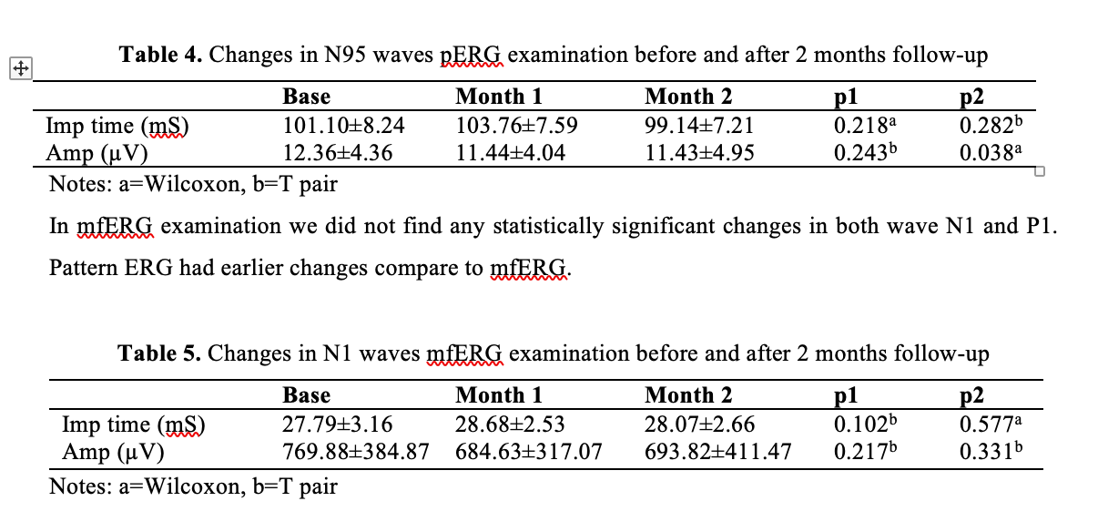
Result: Three studies reported significant thinning of RNFL after ethambutol initiation. Increased RNFL thickness in patients with EON and subclinical EON found in 3 studies. Significant macular GCPIL thinning was noted in 1 study. One study reported shortening of P50 implicit time and reduced N95 wave amplitude in pattern ERG.
Conclusion: Macular GCIPL thinning suggested to be the first pathological changes detected on patients with ethambutol treatment. It can be concomitant with thickening of peripapillary RNFL and followed by peripapillary RNFL thinning. Pattern ERG may reveal abnormality due to retinal ganglion cell (RGC) dysfunction before RGC loss.
Full text article
References
World Health Organization. Global tuberculosis report 2019. WHO; 2019.
Fraunfelder FT, Fraunfelder FW, Chambers WA. Drug-induced ocular side effect, In: Clinical ocular toxicology. Elsevier 2008. p.45-50.
Makunyane P, Mathebula S. Update on ocular toxicity of ethambutol. Afr Vision Eye Health 2016;75(1).a353.
Ezer N, Benedetti A, Darvish-Zargar M, Menzies D. Incidence of ethambutol-related visual impairment during treatment of active tuberculosis. Int J Tuberc Lung Dis 2013;17:447–55.
Lim SA. Ethambutol-associated optic neuropathy. Ann Acad Med 2006;35:274–8.
Lee EJ, Kim SJ, Choung HK, Kim JH, Yu YS. Incidence and clinical features of ethambutol-induced optic neuropathy in Korea. J Neuroophthalmol 2008;28:269–77.
Tsai RK, Lee YH. Reversibility of ethambutol optic neuropathy. J Ocul Pharmacol Ther 1997;13:473–77.
Kumar A, Sandramouli S, Verma L, Tewari HK, Khosla PK. Ocular ethambutol toxicity: is it reversible? J Clin Neuroophthalmol 1993;13:15.
Dialika, Sidik M, Nusanti S, Kekalih A. Correlation between peripapillary retinal nerve fiber layer thickness and visual function changes in patients receiving ethambutol. Medical Journal of Indonesia 2015;24(1).
Jin KW, Lee JY, Rhiu S, Choi DG. Longitudinal evaluation of visual function and structural for detection of subclinical ethambutol-induced optic neuropathy. PloS One 2019;17;14(4):e0215297.
Teng D, Peng CX, Qian HY, Li L, Wang W, Wang JQ, et al. Structural impairment patterns in peripapillary retinal fiber layer and retinal ganglion cell layer in mitochondrial optic neuropathies. Int J Ophthalmol 2018;11(10):1643-8.
Kim KL, Park SP. Visual function test for early detection of ethambutol induced ocular toxicity at the subclinical level. Cutan Ocul Toxicol 2016;35:228-32.
Han J, Byun MK, Lee J, Han SY, Lee JB, Han SH. Longitudinal analysis of retinal nerve fiber layer and ganglion cell-inner plexiform layer thickness in ethambutol- induced optic neuropathy. Graefes Arch Clin Exp Ophthalmol 2015;253: 2293–9.
Kim U, Hwang JM. Early-stage ethambutol optic neuropathy: retinal nerve fiber layer and optical coherence tomography. Eur J Ophthalmol 2009;19: 466–9.
Gumus A, Oner V. Follow up of retinal nerve fiber layer thickness with optic coherence tomography in patients receiving anti-tubercular treatment may reveal early optic neuropathy. Cutan Ocul Toxicol 2015;34: 212–6.
Prakoso BB, Sidik M, Nusanti S, Burhan E, Kekalih A. Changes in pattern and multifocal electroretinogram in tuberculosis patient with ethambutol therapy. Doc Opththalmol 2019;139(1):10.
Kakisu Y, Adachi-Usami E, Mizota A. Pattern electroretinogram and visual evoked cortical potential in ethambutol optic neuropathy. Doc Ophthalmol 1987;67(4):327-34.
Guillet V, Chevrollier A, Cassereau J, Letournel F, Gueguen N, Richard L, et al. Ethambutol-induced optic neuropathy linked to OPA1 mutation and mitochondrial toxicity. Mitochondrion 2010;10:115–24.
Fonkem E, Skordilis MA, Binkley EM, Raymer DS, Epstein A, Arnold WD, et al. Ethambutol toxicity exacerbating the phenotype of CMT2A2. Muscle Nerve 2013;48(1):140–4.
Moon H, Yoon JY, Lim HT, Sung KR. Ganglion cell and inner plexiform layer thickness determined by spectral-domain optical coherence tomography in patients with brain lesions. Br J Ophthalmol 2015;99:329-35:329–35.
Na JH, Sung KR, Baek S, et al. Detection of glaucoma progression by assessment of segmented macular thickness data obtained using spectral-domain optical coherence tomography. Invest Ophthalmol Vis Sci 2012;53:3817–26.
Menon v, Jain D, Saxena R, Sood R. Prospective evaluation of visual function for early detection of ethambutol toxicity. Br J Ophthalmol 2009; 93:1251-4.
Salmon JF, Carmichael TR, Welsh NH. Use of contrast sensitivity measurement in the detection of subclinical ethambutol toxic optic neuropathy. Br J Ophthalmol 1987;71:192–6.
Chai SJ, Foroozan R. Decreased retinal nerve fiber layer thickness detected by optical coherence tomography in patients with ethambutol-induced optic neuropathy. Br J Ophthalmol 2007;91:895-97.
Zoumalan CI, Agarwal M, Sadun AA. Optical coherence tomography can measure axonal loss in patients with ethambutol-induced optic neuropathy. Graefes Arch Cin Exp Ophthalmol 2005;243:410-6.
Kim YK, Hwang JM. Serial retinal nerve fiber layer changes in patients with toxic optic neuropathy associated with antituberculosis pharmacotherapy. J Ocul Pharmacol Ther 2009;25(6):531-5.
Sadun AA, Win PH, Ross-Cisneros FN, Walker SO, Carelli V. Leber’s hereditary optic neuropathy differentially affects smaller axons in the optic nerve. Trans Am Ophthalmol Soc 2000;98:223-32.
Luo X, Frishman LJ. Retinal pathway origins of the pattern electroretinogram (PERG). Investigative ophthalmology & visual science 2011;52(12):8571.
Authors

This work is licensed under a Creative Commons Attribution-NonCommercial-ShareAlike 4.0 International License.