Role of Optical Coherence Tomography Angiography (OCTA) in Anterior Ischemic Optic Neuropathy

Article Sidebar
- anterior ischemic optic neuropathy, optical coherence tomography angiography, peripapillary vessel density
Abstract
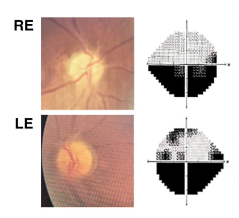
Introduction:Anterior ischemic optic neuropathy (AION) is the most common type of optic neuropathy with symptoms of sudden and painless visual field defect and vision loss. Although evaluating the nonperfusion areas of the vascular ischemia have traditionally been visualized through fluorescein angiography, OCTA has proven to be effective in noninvasively representing the retinal vascular network. This literature review aims to evaluate the quantitaive OCTA assessment of peripapillary vessel density (VD) changes in AION.
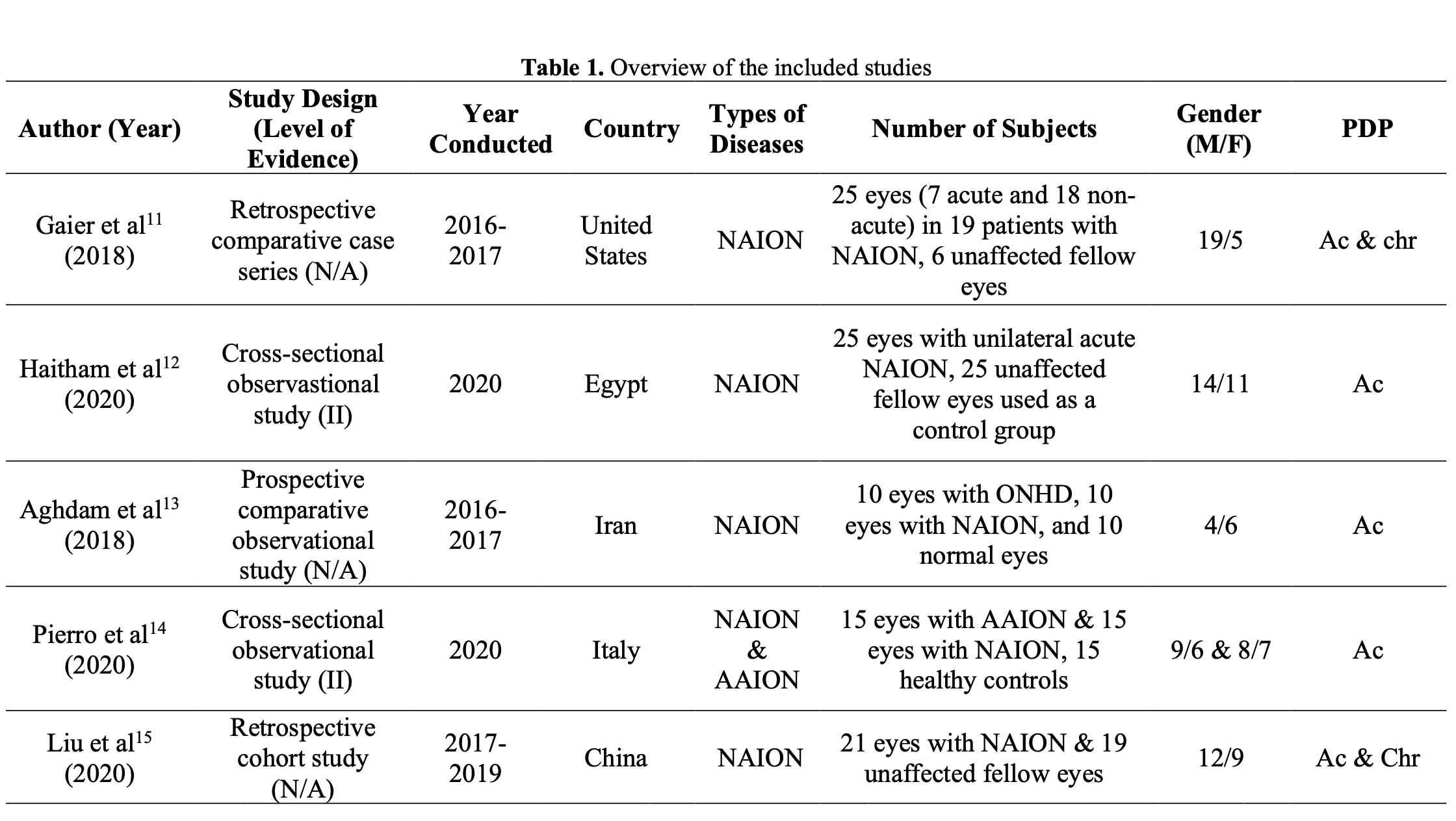
Methods:Literature search was performed in four databases (PubMed, ScienceDirect, ProQuest, and Cochrane Library) from 2018 to 2022 to identify relevant articles. Five studies were included in this review.
Results: All five studies on OCTA findings of NAION eyes reported a reduction in the vessel density of peripapillary capillary plexus when compared to either the healthy control eyes or the fellow unaffected eyes. OCTA reveals vascular changes in both forms, aiding prognosis and treatment. One study comparing NAION and AAION indicates reduced vessel density in NAION and AAION, with more severe abnormalities and reduction of vessel density in AAION.
Conclusion:OCTA can visualize alterations in vascular density in both types of AION, AAION and NAION, with a notably more pronounced reduction in peripapillary vessel density observed in AAION.
Keywords: anterior ischemic optic neuropathy, optical coherence tomography angiography, peripapillary vessel density
Full text article
References
1. Salmon JF. Kanski’s clinical ophthalmology. 9th ed. Oxford: Elsevier Limited; 2020. 755 p.
2. Iorga RE, Moraru A, Ozturk MR, Costin D. The role of optical coherence tomography in optic neuropathies. Rom J Ophthalmol. 2018;62(1):3–14.
3. Patil AD, Biousse V, Newman NJ. Ischemic optic neuropathies: current concepts. Ann Indian Acad Neurol. 2022;25(Suppl. 2):S54-8.
4. Singla K, Agarwal P. Optic ischemia. StatPearls. 2023. p. 1–6.
5. Tan B, Chua J, Lin E, Cheng J, Gan A, Yao X, et al. Quantitative microvascular analysis with wide-field optical coherence tomography angiography in eyes with diabetic retinopathy. JAMA Netw Open. 2020;3(1):1–18.
6. Greig EC, Duker JS, Waheed NK. A practical guide to optical coherence tomography angiography interpretation. Int J Retin Vitr. 2020;6(55):1–17.
7. Jeremy Howick, Chalmers I, Glasziou P, Greenhalgh T, Heneghan C, Liberati A, et al. The Oxford levels of evidence 2 [Internet]. Vol. 1, Oxford Centre for Evidence-Based Medicine. 2011. p. 138–55. Available from: https://www.cebm.ox.ac.uk/resources/levels-of-evidence/ocebm-levels-of-evidence
8. Aumann S, Donner S, Fischer J, Müller F. Optical coherence tomography (OCT): Principle and technical realization. In: Bille J, editor. High Resolution Imaging in Microscopy and Ophthalmology: New Frontiers in Biomedical Optics. Heidelberg: Springer, Cham (CH); 2019. p. 59–85.
9. Raizada K, Margolin E. Non-arteritic anterior ischemic optic neuropathy. StatPearls. 2023. p. 1–5.
10. Blanco-Hernández DMR, Somilleda-Ventura SA, Chávez-Herrera R, Colas-Calvere MG, Lima-Gómez V. Compensatory contribution of retinal larger vessels to perfusion density in diabetics without retinopathy. Sci Rep. 2022;12(1):10–6.
11. Gaier ED, Wang M, Gilbert AL, Rizzo III JF, Cestari DM, Miller JB. Quantitative analysis of optical coherence tomographic angiography (OCT-A) in patients with non-arteritic anterior ischemic optic neuropathy (NAION) corresponds to visual function. PLoS One. 2018 Jun;13(6).
12. Al-Nashar HY, Sahar H. Assessment of peripapillary vessel density in acute non-arteritic anterior ischemic optic neuropathy. Int Ophthalmol. 2020 May;40(5):1269–76.
13. Abri Aghdam K, Ashraf Khorasani M, Soltan Sanjari M, Habibi A, Shenazandi H, Kazemi P, et al. Optical coherence tomography angiography features of optic nerve head drusen and nonarteritic anterior ischemic optic neuropathy. Can J Ophthalmol. 2019;54(4):495–500.
14. Pierro L, Arrigo A, Aragona E, Cavalleri M, Bandello F. Vessel Density and Vessel Tortuosity Quantitative Analysis of Arteritic and Non-arteritic Anterior Ischemic Optic Neuropathies: An Optical Coherence Tomography Angiography Study. J Clin Med. 2020;9(4):1094.
15. Liu J, Chen C, Lu L, Yi Z, Zheng H. Peripapillary and Macular Flow Changes in Nonarteritic Anterior Ischemic Optic Neuropathy (NAION) by Optical Coherence Tomography Angiography (OCT-A). Kurihara T, editor. J Ophthalmol. 2020;2020.
16. Lee Y, Sei KP, Oh Y. Changes in the structure of retinal layers over time in non-arteritic anterior ischaemic optic neuropathy. Eye. 2021;1748–57.
17. Rocholz R, Corvi F, Weichsel J, Schmidt S, Staurenghi G. OCT angiography (OCTA) in retinal diagnostics. In: Bille J, editor. High resolution imaging in microscopy and ophthalmology. Cham: Springer; 2019.
18. Fard MA, Ghahvehchian H, Subramanian PS. Optical coherence tomography in ischemic optic neuropathy. Ann Eye Sci. 2020;5(3):6.
19. Rougier MB, Le Goff M, Korobelnik JF. Optical coherence tomography angiography at the acute phase of optic disc edema. Eye Vis. 2018;5(1):15.
20. Karrabi N, Hooshmandi S, Amirabadi A, Roshandel D, Hassanpour K, Pakravan M. The Role of Optical Coherence Tomography Angiography in Optic Nerve Head Edema: A Narrative Review. Monteiro M, editor. J Ophthalmol. 2022;2022.
Authors
Copyright (c) 2025 Calista Nathasya Gunawan, Syntia Nusanti

This work is licensed under a Creative Commons Attribution-NonCommercial-ShareAlike 4.0 International License.