Non Metallic Anterior Chamber Foreign Body: A Case Report

Article Sidebar
- Anterior Chamber Foreign Bodies (ACFB), IOFB, Open Globe Injury, Penetrating Injury
Abstract
Introduction: Intraocular foreign body (IOFB) is a serious form of open-globe injury that can cause a serious ocular trauma that lead to blindness (10–40% of all open eye injuries). This case report is aimed to report a challenging management of anterior chamber foreign body.
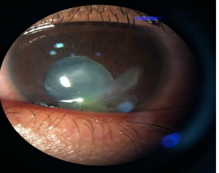
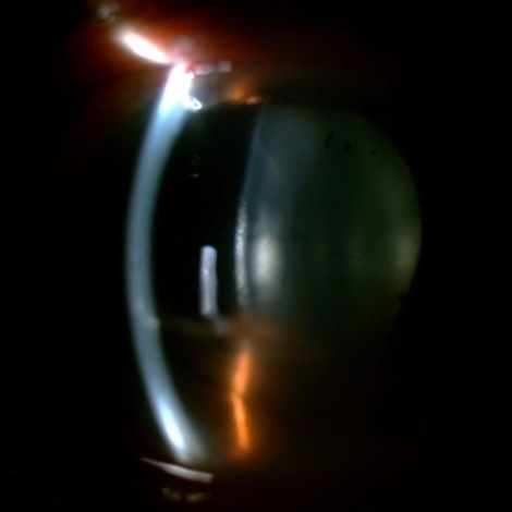
Case Report:A 41 year-old man presented with discomfort on his right eye 4 days prior to visit. The slit lamp biomicroscopic examination revealed inferior anterior chamber foreign body (stone), measuring 5 x 2 mm and 5 mm scar at the area of a full thickness self-sealed corneal laceration. The corneal edema was localized hence it was possible to visualize the foreign body’s entire path through the cornea. The foreign body was removed with forceps from superior limbal incision. Post operative visual acuity was improved and the inflammation was decreased.
Discussion: Management of such cases is not always easy because certain ACFB made of inert materials (stone, plastic, glass, and inert metals such as gold, silver, or platinum) excite minimal inflammation and may remain quiescent for a long period of time. An anterior IOFB is usually associated with a better final BCVA than a posterior IOFB. The self-sealing wounds were limited to the paracentral or peripheral cornea, resulting in no significant astigmatism.
Conclusion: The risk of intraocular foreign body is associated with mechanism of injury and history taking must be accurate. Intraocular foreign bodies must have surgical removal to prevent of ocular inflammation and complication.
Keywords: Anterior Chamber Foreign Bodies (ACFB), IOFB, Open Globe Injury, Penetrating Injury
Full text article
References
Li L, Lu H, Ma K, Li YY, Wang HY, Liu NP. Etiologic causes and epidemiological characteristics of patients with intraocular foreign bodies: retrospective analysis of 1340 cases over ten years. Journal of ophthalmology. 2018 Jan 31;2018.
Chang T, Zhang Y, Zhang K, Zhang X, Wang M, Zeng Y, Zhang M. Epidemiology, clinical characteristics and visual outcomes of patients with intraocular foreign bodies in Southwest China: A 10-year review. Ophthalmic Research. 2020 Nov 13.
Ratanapakorn T, Kongmalai P, Sinawat S, Sanguansak T, Bhoomibunchoo C, Laovirojjanakul W, Yospaiboon Y. Predictors for visual outcomes in eye injuries with intraocular foreign body. Clinical Ophthalmology (Auckland, NZ). 2020;14:4587.
Graffi S, Tiosano B, Ben Cnaan R, Bahir J, Naftali M. Foreign body embedded in anterior chamber angle. Case reports in ophthalmological medicine. 2012 Jan 1;2012.
Yan H, editor. Mechanical ocular trauma: current consensus and controversy. Springer; 2016 Oct 31.
Menon S, Kumar PS, Pai HV. Rare foreign bodies encountered in ophthalmic practice. Journal of Clinical Ophthalmology and Research. 2021 Jan 1;9(1):34.
Al-Tamimi ER. A peculiar case of a retained inert piece of fireworks as an intraocular foreign body in the anterior chamber. Saudi Journal of Ophthalmology. 2014 Jul 1;28(3):225-7.
Su Z, Wang Y, Yi Q, Lin L, Lai K, Ye P, Wang Y, Fang X. Clinical Characteristics and Visual Outcomes in Patients with Intralenticular Foreign Bodies with Self-Sealing Corneal Penetrating Wounds. Journal of Ophthalmology. 2021 Jun 22;2021.
Liu Y, Wang S, Li Y, Gong Q, Su G, Zhao J. Intraocular foreign bodies: clinical characteristics and prognostic factors influencing visual outcome and globe survival in 373 eyes. Journal of ophthalmology. 2019 Feb 13;2019.
Bowen RC, Zhou AX, Bondalapati S, Lawyer TW, Snow KB, Evans PR, Bardsley T, McFarland M, Kliethermes M, Shi D, Mamalis CA. Comparative analysis of the safety and efficacy of intracameral cefuroxime, moxifloxacin and vancomycin at the end of cataract surgery: a meta-analysis. British Journal of Ophthalmology. 2018 Sep 1;102(9):1268-76.
Huang YM, Yan H, Cai JH, Li HB. Removal of intraocular foreign body in anterior chamber angle with prism contact lens and 23-gauge foreign body forceps. International journal of ophthalmology. 2017;10(5):749.
Jastaneiah SS. Long-term corneal complication of retained anterior chamber-angle foreign body. Saudi Journal of Ophthalmology. 2010 Jul 1;24(3):105-8.
Loporchio D, Mukkamala L, Gorukanti K, Zarbin M, Langer P, Bhagat N. Intraocular foreign bodies: a review. Survey of ophthalmology. 2016 Sep 1;61(5):582-96.
Su Z, Wang Y, Yi Q, Lin L, Lai K, Ye P, Wang Y, Fang X. Clinical Characteristics and Visual Outcomes in Patients with Intralenticular Foreign Bodies with Self-Sealing Corneal Penetrating Wounds. Journal of Ophthalmology. 2021 Jun 22;2021.
Li Y. Atlas of ocular trauma. Yan H, editor. Springer Singapore; 2019.
Authors
Copyright (c) 2025 Nabilah Afifah, Nina Handayani , Triana Budi Sulistya

This work is licensed under a Creative Commons Attribution-NonCommercial-ShareAlike 4.0 International License.